NFL stars Russell Wilson (calf muscle tear), Aaron Rodgers (Achilles tendon rupture), Christian McCaffrey (Achilles tendonitis), and Justin Herbert (plantar fasciitis) have all suffered from major orthopedic issues in the last 12 months. On the surface these are all very different orthopedic diagnoses. However, the mechanism by which these injuries occurred is actually quite similar! Furthermore, it’s related to their elite athleticism and the dynamic moves required in a sport like football.
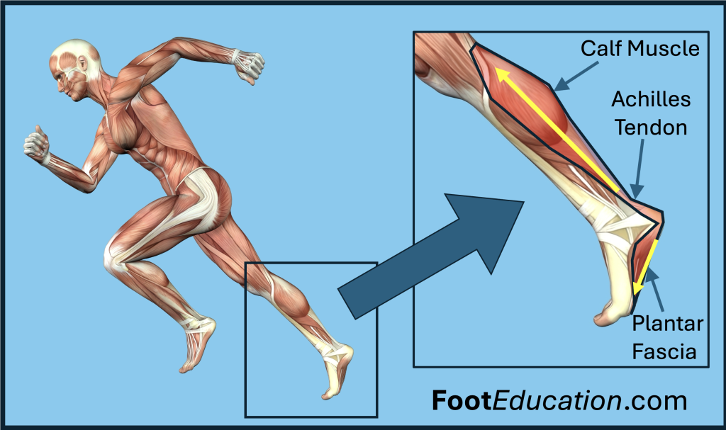
To understand how these various injuries develop, and how they’re interrelated, it is necessary to understand the mechanism by which the forces needed to stand, walk, run, and changed direction are generated in the lower extremity. A review of lower extremity anatomy illustrates that the calf muscle, Achilles tendon, and plantar fascia are all distinctly different structures. However, practically, they function as a unit and can be envisioned as an interconnected whole (Figure 1). A sling of sorts running from behind the knee to the base of the toes. When pushing off during walking or running the calf muscle contracts applying force to the Achilles tendon, which in turn applies force to the heel bone and indirectly to the plantar fascia with the foot acting as a lever to magnify these forces.

The forces going through these interconnected elements are even greater when an elite athlete makes a sudden explosive move to change direction. In this situation the calf muscle is not only contracting to apply force through the other elements, but additionally, the foot is moving upwards immediately prior to changing motion. This means the calf muscle is also lengthening. This is known as an eccentric contraction and the effect of this type of movement is that the internal forces created within all of these structures is many times body weights. The point of all this is that each of these individual anatomical structures (calf muscle, Achilles tendon, and plantar fascia) are subject to considerable force with isolated dynamic movements –and through repetitive action. What determines which specific orthopaedic condition each individual suffers is determined by the location of their particular “anatomic weak link.”
An Achilles tendon rupture occurs when the internal forces generated are greater than the tendon can handle. The Achilles weakens and stiffens with age. When the Achilles tendon ruptures it does so in the same explosive way that an excessively stretched rubber band breaks –a sudden catastrophic failure! This is contrasted with Achilles tendonitis, a condition where individual fibers of the Achilles are microscopically injured, but the overall tendon remains intact. The forces leading to the development of Achilles tendonitis occur from the same mechanism as an Achilles rupture! However, rather than a single catastrophic failure of the tendon repetitive microscopic injuries occur. Imagine a rope swing that is repetitively used over for many years. The rope will tend to develop some fraying while still remaining intact. The body responds to these microscopic injuries to the tendon fibers by sending increased blood and inflammatory mediators to the Achilles. This creates pain, swelling, and dysfunction that is seen in Achilles tendonitis.
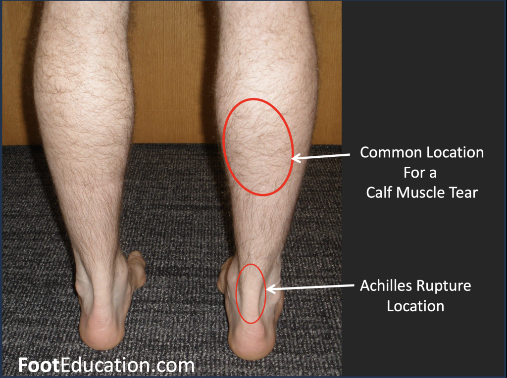
Alternatively, when an individual suffers a calf muscle tear the site of anatomical failure is the calf muscle –usually the inside lower part (Figure 2). The extensive forces the calf muscle is subject to combined with stiffening and weakening with age predisposes an older athlete to suffer a calf tear. This injury is not as catastrophic as an Achilles tendon rupture because much of the calf muscle and it associated fascia usually remains intact. However, a calf tear is very painful, and recovery often takes many weeks before dynamic sporting activities can be resumed.

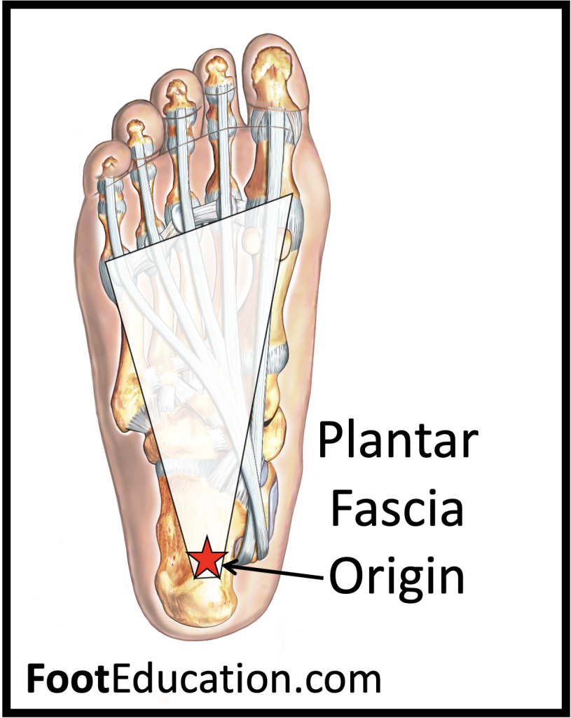
Plantar fasciitis develops from repetitive loading similar to an Achilles tendonitis, albeit with the weak link being the point where the strong plantar fascia inserts into the heel bone (Figure 3). The dynamic forces creating the microscopic injuries in this area are the same force through the anatomical sling that also create the other injuries. The plantar fascia can rupture from this mechanism. However, more commonly the plantar fascia remains intact but suffers microscopic injuries from the repetitive loading. With the plantar fascia remaining intact recovery from plantar fasciitis is usually quicker. Many athletes can play through the condition, although pain symptoms form plantar fasciitis can be quite debilitating.

This interconnected sling of calf muscle – Achilles tendon – heel bone – plantar fasciitis combined with the dynamic forces created by sprinting and sudden change of direction movements in athletes leads to all these different injuries. Same force mechanisms! Different injuries! Furthermore, having a tight calf muscle and explosive muscle strength often allows elite athletes to run faster and make quicker changes in direction –but the price they pay for this athletic advantage is probably an increased risk of suffering one of these injuries -especially as they age.
November 17th, 2024