Ankle Arthroscopy
Indications
An ankle arthroscopy can be used to treat various injuries and disorders of the ankle. These include:
- Synovitis. Synovitis is inflammation of the lining of the ankle joint (synovium). This can cause pain, swelling, and limit movement of the ankle. Synovitis may occur from an injury, arthritis or overuse. Arthroscopy can be used to remove the inflamed synovium.
- Impingement. Impingement of the ankle can occur in the front (anterolateral or anterior impingement) or back of the ankle (posterior ankle impingement). It can cause pain and limit movement of the ankle. Arthroscopy can be used to remove the source of the pain.
- Osteochondral lesion of the talus (a.k.a. Talar OCL or Osteochondritis dessicans). An ankle arthroscopy is often used to assess the Talar defect -and help treat it surgically.
- Loose bodies. Sometimes a loose piece of bone or cartilage can dislodge from within the ankle joint. This piece can be removed with arthroscopy
- Infection. Infections can sometimes occur in the ankle joint either from a previous surgery or from spreading from another place in the body. Arthroscopy may be used to remove infectious tissue and to clean the joint with water to aid in removing the infection.
- Ankle arthritis. Arthroscopy may be used clean bone spurs, and inflamed tissue or loose cartilage associated with ankle arthritis. This does not fix the arthritis, but may potentially help in temporarily improving pain. In very specific situations, arthroscopy may be used to perform an ankle fusion which is one type of surgical treatment for ankle arthritis.
- Ankle instability. Arthroscopy may be used to inspect the ankle joint and even repair ankle instability. This often requires tightening of the ligaments on the outside of the ankle (lateral ligament reconstruction).
- Ankle fractures. Some ankle fractures extend into the ankle joint itself. Ankle arthroscopy may be used to assess for damage to the ankle joint itself, and in some cases help ensure the break is aligned correctly.
Procedure
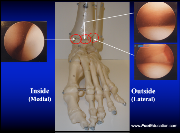
Ankle arthroscopy typically involves two small (~3mm) incisions in the front of the ankle. Through one of the incisions a pencil-sized camera is placed into the ankle. The camera is able to project the live images from the ankle onto a television type monitor (Figure 1). The surgeon is able to then see inside the ankle joint by looking at the images on the monitor. Through the second incision small instruments, which have various purposes (shaving, scraping, biting etc.) can be placed in the ankle. This technique can be used to surgically address many different problems affecting the ankle joint.

Recovery
The recovery plan after an ankle arthroscopy will depend on the type of surgery performed. Usually 1-2 stitches are placed in each incision. These sutures will be removed in the office in about 2 weeks after the wound has healed. After surgery, some patients will be placed in a surgical boot and will be allowed to walk immediately. In other types of surgery a soft cast will be applied and the patient may need to stay off the leg for a period of time. The period of “non-weight-bearing” depends on the type of surgery that was performed, and the surgeon’s post-operative protocol. This time typically ranges from 1-6 weeks. The orthopaedic surgeon will determine the post-operative recovery plan.
Potential General Complications
The complication risks from ankle arthroscopy are very, low about 3.5%, but can include:
- Infection
- Wound Healing Problems
- Blood Vessel Injury
- Nerve Injury
- Breakage of Instruments in the Ankle
- Deep Vein Thrombosis (Blood Clot)
- Pulmonary Embolism (PE)
- Complex Regional Pain Syndrome
- Failure to Resolve ALL Symptoms
Patient Handout: Ankle Arthroscopy
Edited December 2nd, 2023
Previously edited by Paul Juliano, MD, and Sarang Desai, DO
sp/12.2.23