Subtalar Fusion
(Arthrodesis)
Edited by Anthony Van Bergeyk, MD
Watch Video: Subtalar Fusion
Indication
The main indication for a subtalar fusion is to treat painful arthritis in the subtalar joint (the large joint above the heel bone and below the ankle). Arthritis in this joint is commonly seen after heel bone fractures or joint dislocations. Another indication for a subtalar arthrodesis is for patients who need the change to position of the hindfoot in order to distribute load more evenly. For example, a patient with acquired flatfoot deformity, a condition where the heel is offset and load is unevenly distributed, might consider a subtalar fusion.
Procedure
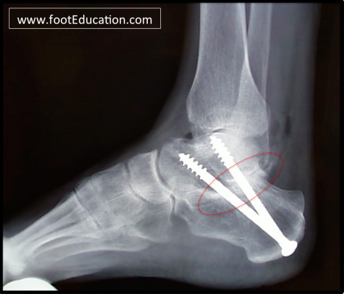
On the lateral (outside front) region of the ankle, a cut is made down to the subtalar joint to expose the joint, particularly the larger posterior portion of the joint, or facet. Once exposed, the joint is ready for fusion. First, the cartilage, or what is left of the cartilage, is removed between the heel bone and the talus. Next, the bone is shaved down to a point that can fuse, or heal together like a fractured bone would heal together. The joint is then placed in its desired position and secured with screws (Figure 1). To increase the chance of the joint fusing together, a bone graft is sometimes done, where a piece of bone is taken from another place in the body (e.g. iliac crest of the pelvic bone) or from a cadaver donor. In some select cases, the subtalar joint can be exposed and fused with small-incision arthroscopic techniques.
Figure 1: Subtalar fusion secured with screws

Recovery
Patients undergoing this type of surgery will typically need about 6-12 weeks for the bone to heal. During this period, the patient is either in a cast or a cast boot, and is non-weight bearing or touch weight-bearing. After the initial non weight-bearing period of 6-8 weeks an x-ray is taken and if healing is progressing well or complete then the patient can begin weight-bearing in a cast boot as tolerated for the next 4-6 weeks and begin physical therapy. Physical therapy exercises include motion of other joints in the foot, strengthening, and gait. After the 10-14 week mark, the patient can transition into a shoe. During the first 5-6 months, patients generally get about 75-80% of recovery; however, it will take about 15-18 months for maximal improvement.
Potential General Complications
- Wound Healing Problems
- Infection
- Deep Vein Thrombosis (Blood Clot)
- Pulmonary Embolism (PE)
- Asymmetric Gait (leading to pain elsewhere)
- Failure to Resolve ALL Symptoms
Potential Specific Complications
- Painful Retain Hardware: Pain may be associated with the screws that are used to secure the joint. About 10-20% of people will need to undergo removal of the screws due to discomfort, once the bones have healed.
- Nerve Injury: Injury to the nerve on the outside of the heel (sural nerve) can occur due to the placement of the incisions. Nerve injury can occur due to retraction, direct injury, or from scarring during the recovery process. If the sural nerve is injured or cut, the patient could end up with numbness or pain along the path of the nerve.
- Nonunion: A nonunion occurs when the fusion site fails to adequately knit together. The nonunion rate is dependent upon: the surgical technique; the patient’s underlying condition; whether the patient smokes cigarettes; and the patient’s compliance with the post-operative non weight-bearing restriction period. The typical nonunion rate is on the order of 5% for the subtalar joint. If a patient does have a nonunion or a delayed union, they may require a longer period of non weight-bearing, and in some instances will require revision surgery.
Edited November 14, 2016
mf/ 3.27.18