Calcaneal Osteotomy
Edited by Matthew Buchanan, MD
Indications
A calcaneal osteotomy is a bone cut (osteotomy) that a surgeon makes across the heel bone (calcaneus). The purpose of a calcaneal osteotomy is to shift the heel bone towards the inside (medial) or outside (lateral). If perfectly aligned, your heel bone should be directly underneath your shin bone (tibia). Some patients have a heel bone that is shifted or tilted too far to the inside or outside of their shin bone. This abnormal alignment can have a negative effect on the function of the foot and ankle but also the knee and hip. The goal of a calcaneal osteotomy is to correct the alignment of the heel bone so that it lies directly underneath the shin bone.
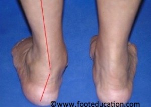
A calcaneal osteotomy is indicated for patients who have failed a non-operative treatment program for their heel alignment abnormality. Depending on the alignment abnormality, the heel (calcaneus) may be shifted towards the inside (medializing calcaneal osteotomy) or the outside (lateralizing calcaneal osteotomy). For example, a patient with a flat foot (pes planus or acquired adult flatfoot deformity), will have their heel abnormally shifted too far to the outside (Figure 1). This alters the way load is distributed through the heel. Individuals with a flat foot typically have pain and tearing of the soft tissues (ligaments and tendons) along the inside of the foot and ankle.
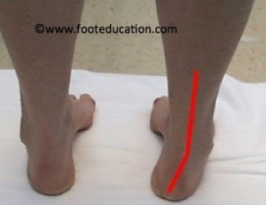
A patient with a high arch (pes cavus) will have the heel abnormally shifted too far to the inside (Figure 2). This may create pain, stretching out, and even tearing of the soft tissues (lateral ankle ligaments and peroneal tendons) on the outside of the foot and ankle. This stretching out of ligaments increases the likelihood of ankle sprains and may result in chronic ankle instability. In addition, a heel that is abnormally shifted too far to the inside or outside can alter the weight bearing forces through ankle joint, causing abnormal cartilage wearing (arthritis).
Figure 1: Left heel abnormally shifted to the outside in flatfoot deformity

Figure 2: Right heel abnormally shifted to the inside in a high arched foot.

Procedure
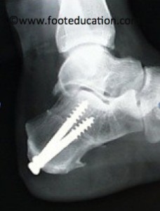
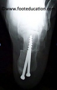
A skin incision is made on the outer aspect of the heel bone. Your surgeon may choose to perform this procedure through a traditional incision or a minimally invasive (smaller) incision. The soft tissues (nerves and vessels) in the area are protected and a fine cut is made across the heel bone. The back part of the heel bone is then shifted towards either the inside or outside. Once shifted, the bone is fixed in place with the use of one or two screws (Figure 3).
Figure 3: Calcaneal Osteotomy (Side and Bottom View)


Recovery
0-6 weeks Post-Surgery
Patients undergoing this type of surgery will typically need about 6 weeks for the bone to heal. During this period, the patient is either in a cast or a cast boot and remains non-weight bearing or touch weight-bearing.
6-12 weeks Post-Surgery
During weeks 6-12, patients can begin weight bearing in the CAM Boot. This is a gradual process that incrementally increases the amount of force placed through the heel bone. Once the patient achieves full weight bearing in the boot without pain, they begin to switch over to a supportive shoe and slowly increase the number of steps that they take each day. It can be up to 12 weeks before a patient is in a shoe for 100% of the day. Supervised physical therapy is often recommended to assist with the post-surgical rehabilitation.
Calcaneal osteotomies are often combined with other procedures, such as tendon transfers, so the actual recovery time may vary depending on the procedures that are performed. In general, 75-80% of the recovery occurs within the first 6 months. It takes 12 months or more for the musles, tendons, and bones to recover 100%.
Potential Complications
- Wound Healing Problems
- Wound Infection
- Deep Vein Thrombosis (DVT)
- Pulmonary embolism (PE)
- Complex Regional Pain Syndrome (CRPS)
- Asymmetric Gait
Specific potential complications
Complications specific to this surgery include:
- Sural nerve and medial calcaneal nerve injury: The incision over the outer aspect of the heel bone and shifting of the actual bone can place nerves at risk. The sural nerve is located in close proximity to the incision and can be injured during the surgical approach to the bone. The medial calcaneal nerve is on the inside of the heel bone and can be injured during the bone cut or during the shifting of the bone. If these nerves are injured or cut, the patient could end up with numbness or pain along the path of the nerve
- Painful Hardware: Another potential complication with this procedure is having pain associated with the screws that are used to secure the heel. About 10-20% of people will need to undergo removal of the screws due to discomfort. Screws are not removed until the bone has completely healed.
Previously Edited by Vinod Panchbhavi MD and Michael Castro, DO
Edited October 29. 2019
mf/ 11.5.18