Plantar Fibroma
Edited by Michael Castro, DO
Summary
A plantar fibroma is the most common reason for a lump to develop on the arch of the foot. These are often small (less than half an inch), but can grow steadily to reach sizes of 2 inches or more. They may occur as a single lesion or they may present as multiple lesions, typically along the medial border of the plantar fascia. They are thought to arise from stress or repetitive trauma to the tissue, but may also have a genetic component. They are benign and are composed of dense, fibrous tissue found in the ligaments. Rapid-growing and multi-planar fibromas are considered plantar fibromatosis.
Many patients with plantar fibromas do not have any symptoms and when they do, it is often only a vague discomfort from “walking on a lump.” These masses can be treated non-surgically by reducing the stress on the plantar fascia or with a custom or soft accommodating prefabricated (over the counter) orthotic. However, if they grow larger or are a persistent source of discomfort, they should be removed surgically.
History
Patients with plantar fibromas will usually report that they “just noticed the mass one day.” Occasionally they will have a history of a similar mass in the palm (Dupytren’s Contracture). As the mass gets larger, they may report increasing discomfort with prolonged standing.
The first thing to look for however is a foreign body reaction from previous penetrating trauma, which is the most common cause of benign tumor of the sole (especially among diabetic patients).
Second, although uncommon a malignant synovial sarcoma may cause a fast growing and painful lump on the sole of the foot.
Physical Examination
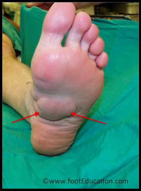
Plantar fibromas lumps that are usually located on the inside of the arch of the foot (Figure 1). The lump usually feels smooth and rubbery. They are not usually tender to touch, although they may become irritated with prolonged walking. Testing ankle motion and tension of the calf muscles is important and may identify the source of the irritation. Like plantar fasciitis, the stress applied to the fascia can be associated with calf muscle tightness causing early heel rise.
Figure 1: Large Plantar Fibroma in classic location on the sole of the foot

Imaging Studies
X-rays
X-rays are often negative, although if the x-rays are taken in such a way as to highlight the soft-tissues, an outline of a mass may be seen. Calcifications are looked for since it is a sign of malign tumor and would require an MRI.
MRI
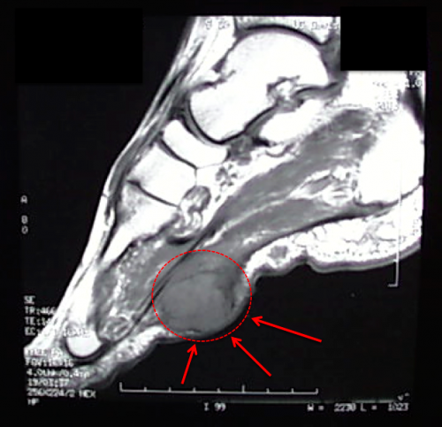
An MRI will show a smooth, consistent (homogenous) mass that is affiliated with the plantar fascia (Figure 2). An MRI will confirm the diagnosis and allow differentiation of other causes of masses in the foot, such as lipomas, ganglions, neuromas, herniations of the plantar fasica, and tumors (very rare).
Figure 2: MRI of a large plantar fibroma

Treatment
Non-Operative Treatment
Plantar fibromas are typically treated non-operatively, until they cause discomfort to the patient on a regular basis. Non-operative treatment includes:
- anti-inflammatory medications
- calf muscle stretching
- shoe modifications
- the use of a soft-accommodating or well molded custom-made shoe insert
Injectable Medications
Much of the conservative treatment lacks direct supporting evidence-based medicine and studies to support its efficacy, many of the modalities available derive from Dupuytren’s disease conservative treatment. Injections can shrink the plantar fibroma and reduce the symptomatology whereas excision surgery has a high recurrence rate and increased the potential for wound healing issues.
Injection is mainly of corticosteroid (15 mg to 30 mg of triamcinolone acetonide in each nodule). A series of three to five injections with each injection spaced three to six weeks apart. However, 50 percent of the patients experienced recurrence within one to three years after their last injection, necessitating one or more additional injections. Secondary effects are transient depigmentation or temporary subcutaneous fat atrophy at the injection site. On the treatment horizon is the possible injection of collagenase.
Operative Treatment
Operative treatment is typically avoided due to both the high recurrence rate and the difficulty with obtaining clear margins. Consideration should be given to lengthening of the gastrocnemius tendon (gastrocnemius recession) if the muscle is identified as a contributor. Gastrocnemius recession is performed through a small incision along the inside of the calf. The procedure and follow-up physical therapy can result in improved ankle motion. The improvement in ankle motion decreases the stress on the plantar fascia. The “bump” may not disappear but there is often significant pain relief. The benefit being no time off the foot with the return to normal activity in 4 to 8 weeks.
An alternative treatment involves excising (cutting out) the mass. This is typically done through an incision in line with the length of the foot. The mass is then sent to be examined by the pathologist to confirm the diagnosis. Recovery and return to unrestricted activity and shoewear is in the one-to-two-month range.
Potential complications of surgery
Complications associated with gastrocnemius recession are few.
- Infection: Infection at the surgical site typically occurs in less than 2% of patients.
- Nerve Injury: The sural nerve runs along the backside of the gastrocnemius and can be irritated by swelling.
- Blood clot (DVT): Risk of a blood clot is rare with immediate post-operative weight bearing.
Surgery to remove a plantar fibroma can be associated with the usual array of potential complications, including:
- Recurrence: These lesions typically involve the overlying skin and dermis, as well as the plantar fascia, and obtaining clean margins is difficult. As a result, they have a high recurrence rate and in many cases, recur rapidly. Some authors advocate complete removal of the plantar fascia to reduce the rate of recurrence. Surgical margins should approach 2 cm. The recurrence rate is significantly higher for plantar fibromatosis and in revision cases. Adjuvant radiation therapy may be indicated to reduce the incidence of recurrence.
- Wound healing problems: Wound complications are surprisingly common in this type of surgery, as the lesions typically involve the overlying epidermis and dermis (3-30+% depending on the size of the mass). Patients should be aware that skin grafting may be necessary. The larger the mass, the greater the likelihood of a wound problem. A painful scar is always a possibility with surgery on the sole of the foot, although it happens uncommonly.
- Infection: Usually occurs in 2-5% of cases, although it may be higher if a wound problem develops
- Nerve injury: 2-5% of patients may have injury or irritation to the nerve on the inside of the sole of the foot (medial branch of the plantar nerve).
- Blood clot (DVT): These are quite uncommon unless there is a specific risk factor for blood clotting.
- Pulmonary Embolism: Potentially very serious, but very uncommon unless there is a specific risk factor for blood clotting
Edited on August 21, 2017
(Originally edited by Jean Brilhaut, MD and Matt Buchanan, MD)
mf/ 5.21.18