
Cuboid Impaction (Nutcracker) Fracture
Summary
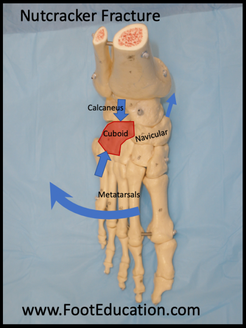
A cuboid impaction fracture, also referred to as a nutcracker fracture, is an uncommon fracture of the cuboid bone on the outside of the foot (Figure 1). This injury occurs in the setting of a forceful outward (eversion-abduction) or downward (plantarflexed) movement of the foot. This leads to a crushing of the cuboid bone between the bones on either side of it (calcaneus and 4th and 5th metatarsal bases). In a classic “Nutcracker fracture” there is also a pulling off type (avulsion) fracture of the navicular bone on the inside (medial) foot. Patients present with pain and swelling of the foot, particularly the outer side. Diagnosis is made based on x-ray findings, sometimes supplemented by a CT scan. Treatment involves pain management and immobilization. Surgery to reposition (reduce) the fracture(s) is indicated when the fracture(s) are displaced. Recovery from a cuboid impaction fracture can be prolonged.
Figure 1: Nutcracker Fracture (Cuboid Impaction) Mechanism
Clinical Presentation
The nutcracker fracture can be observed in ballet dancers or horseback riders, whose feet are commonly in an “en pointe” (pointed/plantar flexed) position. Patients will present with a history of acute trauma to a plantarflexed foot. Patients will often experience pain on the outer (lateral) side of the foot, and have difficulty with weight bearing on the injured foot.
Physical Exam
Diagnosis of a nutcracker fracture during physical examination is challenging because nutcracker fractures are uncommon and the mechanism of injury is similar to that of a Lisfranc injury. Physical examination will commonly reveal pain and swelling around the cuboid – on the outside /lateral aspect of the foot. There is often pain on the inside (medial) of the foot as the pull of the posterior tibial tendon can cause a section of the navicular bone to be pulled off (avulsed). A severe displaced nutcracker fracture will be associated with an observable deformity of the lateral side of the foot.
Imaging
X-ray
X-rays are commonly used to identify and characterize cuboid injuries. A nutcracker fracture of the cuboid is best visualized on the oblique view of an x-ray. This view reveals the relationships between the cuboid bone and the 4th and 5th metatarsal bones located in front (anterior side) of the cuboid. The oblique view may also be used to identify the length of the lateral part of the foot, which can be expected to change when the cuboid bone is crushed.
CT and MRI
Advanced imaging such as a Computed Tomography (CT) or Magnetic Resonance Imaging (MRI) can be used to better understand the fracture pattern involving the cuboid and identify any associated injury to other bones, ligaments, or soft tissue.
Treatment
Treatment of a nutcracker fracture is dependent on the extent of the injury. An undisplaced or minimally displaced cuboid impaction fracture can be treated with rest, ice, elevation, and immobilization in a cast or boot. More severe nutcracker fractures are treated surgically. Surgery aims to reposition the broken bones back to their pre-fracture position (fracture reduction) and stabilize then there usually with screws and a plate (internal fixation). Bone healing usually takes 6-10 weeks so patients require a prolonged period of non-weight-bearing or minimal weightbearing to facilitate bone healing. After adequate bone healing has been achieved physical therapy to regain motion and strength is often beneficial. Progression back to physical activity such as jogging and running can take many months.
Edited June 20th, 2020