

Ligaments of the Foot and Ankle Overview
Definition
Ligaments are strong connective tissue composed of fibrous tissues. They connect bones to other bones, and are extremely important in stabilizing joints.
The ligaments of the foot and ankle can be divided into groups including:
1. Ligaments Stabilizing the Ankle Joint
- The anterior Talo-fibular ligament (outside or lateral ankle joint)
- The Calcaneo-fibular ligament (outside or lateral ankle joint)
- The posterior Talo-fibular ligament (outside or lateral ankle joint)
- The Deltoid ligament (inside or medial ankle joint)
2. Ligaments of the Upper Ankle (Holding the tibia and fibula together)
- The Anterior Inferior Tibiofibular Ligament
- The Interosseous Ligament
3. Ligaments of the Subtalar Joint
- The Cervical Ligament
- The Lisfranc Ligaments
- The Intermetatarsal Ligaments
- The joint capsule of the Great Toe
1. Ligaments Stabilizing the Ankle Joint
The Anterior Talo-Fibular Ligament (Figure 1)
The anterior talo-fibular ligament is often thought of as the “ankle sprain ligament”, because it is the one that is most often damaged when the ankle is rolled. It runs down from the fibula and to the outer front portion of the ankle, in order to connect to the neck of the talus. This ligament functions to stabilize the ankle joint and keeps it from rotating inward too much, which is what happens in a common ankle sprain. The ligament itself is very broad, more like a sheet than a rope, and when it is sprained, it tends to tear part of the way rather than into two pieces. Another possibility is that it becomes stretched out in places, which makes the ankle joint looser than normal, potentially leading to symptomatic ankle instability.
Figure 1: Lateral Ankle Ligaments
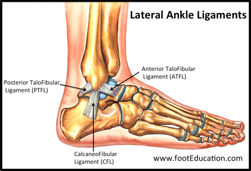

The Calcaneo-Fibular Ligament (Figure 1)
The calcaneo-fibular ligament is almost the “baby brother” of the anterior talo-fibular ligament. It starts at the tip of the fibula and runs along the outside of the ankle, into the calcaneous. It is more rope-like in structure than the anterior talo-fibular ligament, and less susceptible to damage, although it can be damaged during a severe ankle sprain.
Posterior Talo-Fibular Ligament (Figure 1)
The posterior talo-fibular ligament runs from the back lower part of the fibula and into the outer back portion of the calcaneous. This ligament functions to stabilize the ankle joint and subtalar joint. Injuries are rare to this ligament, unless there is an ankle dislocation or subluxation.
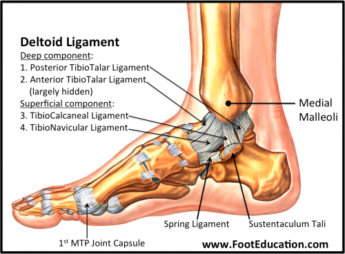
The Deltoid Ligament (Figure 2)
The deltoid ligament is a fan shaped band of connective tissue on theinside of the ankle. It runs from the medial malleolus (the bottom portion of the tibia) and down into the talus and calcaneous. The deeper branch of the ligament is securely fastened in the talus, while the more superficial, broader aspect runs into the calcaneous. This ligament is extremely strong and stabilizes the inside of the ankle. It can be torn, but it takes tremendous force. Tears of this ligament are most common when the ankle undergoes an unstable ankle fracture. Like the anterior talo-fibular ligament, the deltoid is rarely torn completely and tears tend to resemble a torn or stretched sheet.
Figure 2: Medial Deltoid Ligament

2. Ligaments of the Upper Ankle (Holding the tibia and fibula together)
Anterior Inferior Tibial Fibular Ligament (Figure 3)
The anterior inferior tibial fibular ligament is commonly known as the “high ankle sprain ligament”. It is positioned on the lower front of the ankle and helps keep the tibia and fibula together. Injuries to this ligament occur when the foot is stuck on the ground and rotated inwardly. This injury can be very painful and can scar, which can cause irritation to the outside of the ankle joint, a condition known as anteral-lateral ankle impingement.
Figure 3: Ligaments of the Upper Ankle
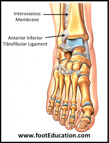

The Interosseous Membrane (Figure 3)
The interosseous membrane is composed of strong fibrous tissue and runs along the tibia and fibula, and keeps the two bones moving as one unit. It can be torn from a type of unstable ankle fracture, known as a Weber Type C ankle fracture, in which the tibia and fibula have to be torn apart.
3. Ligaments of the Subtalar Joint
The Cervical Ligament
Also known as the subtalar ligament, this ligament connects the calcaneous to the talus in front of the subtalar joint. It keeps the calcaneous in place relative to the subtalar joint, so the two bones do not move completely independently of one another.
4. The Ligaments of the Foot
Lisfranc Ligaments (Figure 4)
The Lisfranc ligaments stabilize the small bones of the mid-foot, as well as the transverse tarsal joint. They are stronger across the plantar (sole) of the foot than on the dorsal (top) aspect, though they are very strong in either case. These ligaments were described by the Napoleonic era surgeon Jacques Lisfranc, who noticed that when people fell from their horses but got their foot stuck in the stirrup, they dislocated portions of the mid-foot. These dislocations (Lisfranc Injuries) are very problematic, and took a long time to heal.
Figure 4: Plantar Ligaments
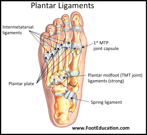

The Inter-Metatarsal Ligaments (Figure 4)
These ligaments run between the metatarsal bones at the base of the toes. They connect the neck region of each metatarsal to the one next to it and bind them together. This keeps the metatarsals moving in sync. While it is possible to tear these ligaments, it is also possible for them to irritate the digital nerve as it crosses the ligaments, potentially leading to a Morton’s neuroma.
The Joint Capsule of the Big Toe (Figure 4)
The connective tissue of this ligament takes the form of a capsule. It goes from the inner portion of the first metatarsal head and stretches to the distal phalanx on the inside. This allows this ligament to stabilize the great toe on the inside. In the situation where a person develops a bunion, this band gets stretched out, and the toe changes position because of this.
Edited October 17, 2015
mf/ 9.4.18