

Posterior Ankle Impingement
(Os Trigonum Syndrome)
Edited by Kalpesh Shah, FRCS
Summary
Posterior Ankle Impingement is characterized by pain behind the ankle joint. Symptoms are exacerbated by pointing the toes in a forced position. Posterior ankle impingement is differentiated from insertional Achilles tendonitis and retrocalcaneal bursitis, in that these conditions are more closely associated with the attachment of the Achilles tendon into the heel bone (calcaneus). Treatment is usually non-surgical, including rest, activity modification, anti-inflammatory medications, and occasionally a corticosteroid injection. If there is a mechanical impingement or a large extra bone behind the ankle (os trigonum), treatment may include removal of the offending bone spur or bone fragment.
Clinical Presentation
Pain behind the ankle is the primary symptom of posterior ankle impingement. Symptoms are usually aggravated by pointing the toes toward the floor or pointing the toes like a ballerina (forced plantar flexion). This condition may be precipitated by an acute injury and/or may be associated with an extra bone fragment (os trigonum) at the back of the ankle.
Physical Examination
Pain will be localized behind the ankle joint. There may also be some swelling associated. Pointing the toes in a forced position will aggravate symptoms. Sensation and movement is usually normal. Occasionally, the pain will be such that moving the big toe will aggravate symptoms, such as the tendon that runs behind the ankle joint.
Imaging studies
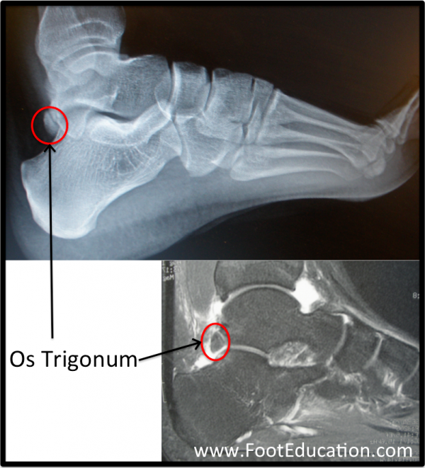
Plain x-rays of the foot and ankle from the side may demonstrate an extra fragment of bone extending from the back of the talus (os trigonum). This is quite a common finding on x-ray, although most os trigonums are asymptomatic. Other sources of pain leading to posterior ankle impingement include bone spurs, or evidence of arthritic changes near the ankle or subtalar joint. For many patients with posterior ankle impingement, the x-rays will be normal.
A bone scan may be used as a non-specific means of localizing the inflamed and irritated area. An MRI can give more detailed resolution to the bony and soft tissue structures in the region.
Figure 1: Os Trigonum (circled in red) on plain ankle x-ray (top) and MRI (bottom).

Treatment
Non-Operative Treatment
If the condition is not related to a mechanical irritation, such as a loose body or a bone spur, the condition can usually be treated successfully without surgery. Rest and relative immobilization, followed by a period of gradually increasing activity, can be very helpful. Occasionally, a steroid injection into the ankle joint may be beneficial.
Surgical Treatment
If a large bone spur, loose body, or symptomatic os trigonum is present and this fails non-operative management, then surgery may be employed to “clean out” this area (debridement). This is often done through an incision on the back outside part of the ankle, but in some instances, it may be performed arthroscopically. If a bone spur or os trigonum is removed, then the ankle is usually not destabilized and weight bearing can be commenced immediately after surgery. There is usually persistent swelling and discomfort after the surgery so limiting activities is required until these symptoms settle.
Previously Edited by Paul Juliano, MD
Edited on October 30, 2018
mf/ 6.4.18